

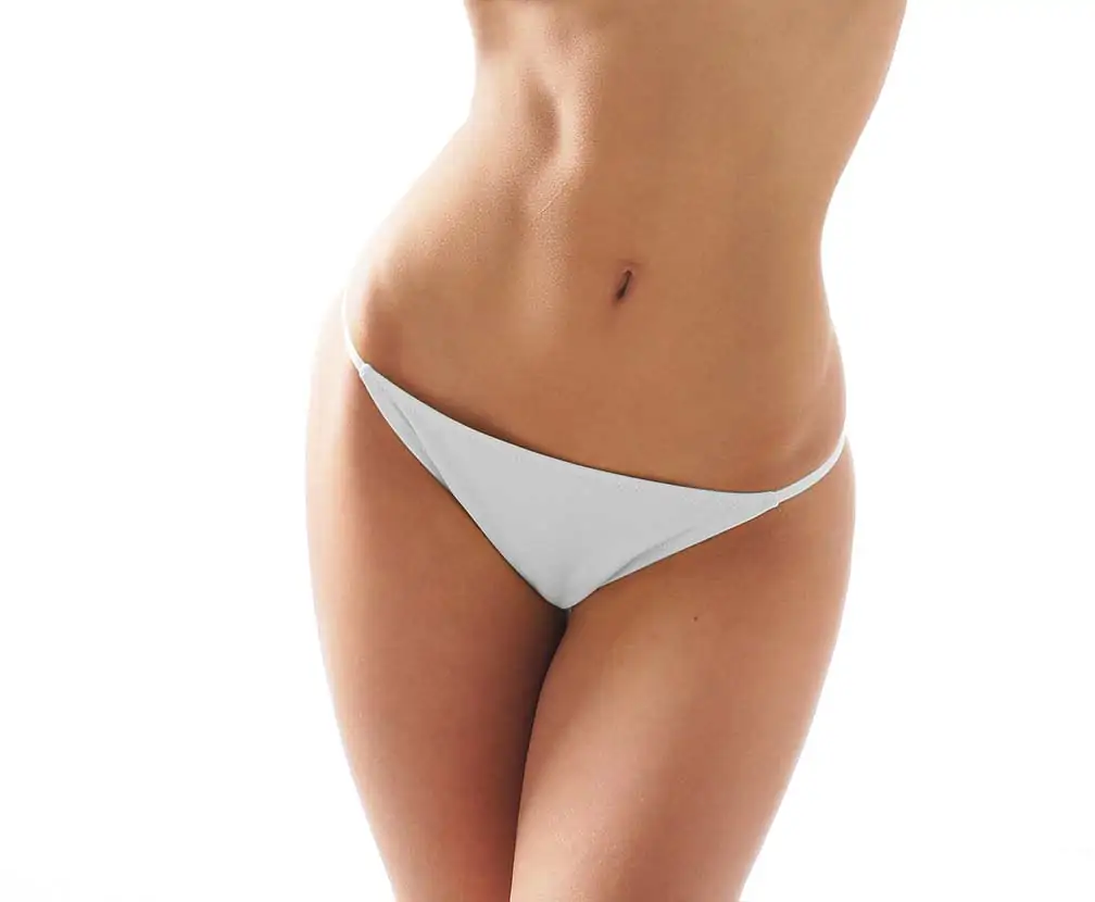
Mommy makeovers help thousands of women every year to address the physical changes of motherhood. Surgeries can be tailored exactly to fit the needs of the patient. They may include any combination of abdominoplasty (tummy tuck), breast augmentation, labiaplasty, liposuction, and a Brazilian Butt Lift (BBL). What’s more, a mommy makeover can renew confidence often lost as the body recovers from the stresses of pregnancy, childbirth, and breastfeeding. (1)
Every woman deserves to feel comfortable in their own skin. That’s why board-certified plastic surgeon Dr. Steven Wallach provides an alluring selection of body-contouring procedures, to help you address pregnancy-related changes on your own terms. In the midst of taking on one of life’s most challenging roles, reclaim a sense of self once more. Call our Manhattan office at (212) 861-6400 or fill out our inquiry form to book your personal consultation with Dr. Wallach to discuss the aesthetic transformation you deserve!
Contents
showLife After Childbirth

The impact of motherhood on the body is often glossed over. Your motherly instincts help you provide for your infant, but your needs are equally important. Over the course of nine months, and well after, your mind and body change immeasurably. And, after giving birth, you may yearn for the pre-pregnancy body you once knew. Unexpected, unwanted, and long-lasting changes from pregnancy can be distressing and uncomfortable, not to mention embarrassing!
Discouragement can set in when an intentionally healthy diet coupled with regular exercise doesn’t tighten excess skin or shed the excess pounds. Nursing can also cause one’s breasts to change from the constant tugging, pushing, and pulling. Additionally, excess skin on one’s abdomen may lack elasticity from carrying a child to term. You may feel a cadre of emotions: frustration, fear, love, and sadness. And as your body and mind recalibrate to a new normal, your self-confidence may also suffer. Dr. Wallach can help you reclaim your mind and body, and recultivate your sense of self and autonomy.
About Mommy Makeovers
The mommy makeover is one of the most popular aesthetic surgeries. It is widely recognized as a safe procedure, with high patient satisfaction rates. (2) This is because no two mommy makeovers are exactly the same. Rather than one-size-fits-all, the surgical services are customized to you. You may want to fix extra skin drooping around your abdomen, remove the sunken appearance of your navel, or deal with overstretched, stubborn “flab” elsewhere. Perhaps your breasts have lost some of their natural lift from nursing as you have decided to place your child’s health first. Whatever your concern, Dr. Wallach can help. As you lay the foundations for your young child to survive and thrive, he can help your body readjust via your unique treatment plan. Your mommy makeover may include but is not limited to: (3)
LIPOSUCTION
Changes in your metabolism during pregnancy can alter fat deposits. To prepare for childbirth, the body stores extra fat on the abdomen, hips, and thighs. This excess tissue may be difficult to remove through diet and exercise alone. Liposuction can help to remove this fat and may be performed in conjunction with a tummy tuck to further contour your abdomen.
TUMMY TUCK
Also called an abdominoplasty, a tummy tuck removes extra skin and fat to tighten and firm a sagging abdominal profile. It can also restore abdominal muscles that are weakened or separated during pregnancy, a condition known as diastasis rectus. Mild to severe diastasis recti can be repaired through abdominoplasty. By suturing the muscle fibers closer to the midline, Dr. Wallach can provide support for a stronger core. A well-hidden hip-to-hip incision means you can achieve a flatter post-pregnancy abdomen, concealing the scar in the bikini line.
BREAST REDUCTION OR AUGMENTATION
Breast reduction or breast augmentation may not be essential components of some mommy makeovers. However, a woman whose cup size is naturally larger may decide to decrease her chest size, likely for comfort and functional reasons. A woman whose cup size is smaller, or whose breasts have decreased in size following breastfeeding, may choose breast augmentation. Dr. Wallach offers augmentation with implants, or via fat transfer from a donor area.
Breast Lift
Many women in their 30s and 40s opt for a breast lift, or mastopexy, after childbirth. Why? A mother’s breasts will fill with milk whether or not she breastfeeds. During this time, a woman’s cup size increases up to a few sizes. Once a child stops breastfeeding, the breasts will contract to their original size and can eventually sag. A breast lift helps to restore a woman’s pre-pregnancy breast projection, raising them to a higher, more youthful-looking chest position.
VAGINAL REJUVENATION
Vaginal rejuvenation can take many forms. Many women notice their labia minora, the inner labia, change shape following vaginal birth. They may also notice tissue changes elsewhere in the vulva. Loose skin may cause chaffing, and intercourse may be uncomfortable. Dr. Wallach offers labiaplasty, labia majora reduction, and mons pubis reduction. Once healed, patients can share intimate moments more comfortably and exercise with confidence.
BRAZILIAN BUTT LIFT
Like other post-pregnancy body areas, a woman’s buttocks may also sag, particularly if she gained weight in this area during pregnancy, then lost it rapidly. A BBL pairs well with liposuction since excess fat harvested by liposuction can be placed in the buttocks for a fuller appearance. For those who do not opt for fat transfer, he offers solid silicone implant buttock augmentation.
Benefits of a Mommy Makeover
Every patient aims to achieve different goals through a mommy makeover. Some of these may include:
- Reduction of stubborn abdominal fat
- Removal of excess skin around the abdomen
- Improvement of stretched nipples and areolas
- Reestablished breast symmetry and profile
- Reshaped labia for improved comfort
- Improvement in clothing fit
- Increased body confidence
Candidates for a Mommy Makeover
How do you know if a mommy makeover is right for you? As it’s not weight loss surgery, good candidates for a mommy makeover:
- Want to remove excess post-pregnancy skin and fat surgically
- Are at or close to their ideal weight
- Have no serious medical conditions
- Have finished building their family
- Have realistic expectations for recovery and results
Should you undergo a mommy makeover and become pregnant, another pregnancy may undo the results of your surgical procedures. Choosing to get a mommy makeover once you are done having children means that the effects of removing excess fat and tightening skin will have a longer-lasting impact. Additionally, as a mommy makeover is not a weight loss procedure, patients should not seek this kind of surgery to lose weight. Lastly, the motivation for your mommy makeover should not come solely from society or a person in your life. The deeper desire to reclaim yourself inside out, to reclaim a body that will naturally be impacted by pregnancy should come from you, and you alone.
Personal Consultation
Tell board-certified plastic surgeon Dr. Wallach what you would like to change about your post-pregnancy body. By booking a personal consultation at his Manhattan office, you can work with him to take the first steps toward designing your unique treatment plan. Arrange your mommy makeover today by contacting us at (212) 861-6400.
What to Expect
Dr. Wallach will give you detailed preoperative instructions before your surgery. These include altering the medications and dietary supplements you take, and stopping smoking entirely if you’re a smoker. The finer details of your procedure will vary depending on your treatment plan.
A mommy makeover is major cosmetic surgery. Patients should be prepared to follow their aftercare instructions carefully. These may include:
- Having an adult on hand 24 hours after the procedure to assist with recovery
- Having someone drive you home after your procedure
- Taking at least two weeks off of work to optimize healing
- Taking pain medication exactly as prescribed
- Avoiding driving for at least one week after surgery
- Sleeping on their back or side with elevated upper body and feet
Dr. Wallach will prescribe pain relief medication for your comfort. Everybody heals uniquely, but patients should expect up to three months for a full recovery with swelling for six weeks or more post-surgery.
Cost of a Mommy Makeover in Manhattan
A mommy makeover constitutes a long-term investment in your appearance and quality of life. The procedure may not be inexpensive, but our patients feel that the results are worth the investment! Please find more information on our financing options on our website.
Restore your body and confidence to the form you knew pre-pregnancy. Call Dr. Wallach at (212) 861-6400 today to learn more.
Read Dr. Wallach’s blog for more information about the mommy makeover and his many other plastic surgery services in New York.
References
- Carniol, E.T. & Carniol, P.J. (2010). The “Mommy Makeover” Package. AMA Journal of Ethics, 12(5), 363-366. https://journalofethics.ama-assn.org/article/mommy-makeover-package/2010-05.
- Vidal, P, Berner, J.P., & Will, P.A. “Managing Complications In Abdominoplasty: A Literature Review. Archives Of Plastic Surgery. 44(5), 457-468. https://www.ncbi.nlm.nih.gov/pmc/articles/PMC5621815/.
- Nahabedian M. Management Strategies for Diastasis Recti. Seminars in Plastic Surgery. 2018;32(03):147-154. https://www.ncbi.nlm.nih.gov/pmc/articles/PMC6057788/


 reviews
  call
call  gallery
  email
email